
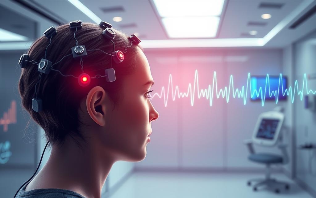
Electroencephalography is a key part of modern neurological diagnostics. It lets doctors see how the brain works. This method uses electrodes on the scalp to measure electrical signals.
The results show brain wave patterns. Experts study these to find out if the brain is working right. This helps them understand different brain conditions.
The Mayo Clinic says EEG is a main tool for finding epilepsy and other brain problems. It’s painless and gives important information for diagnosis.
This technology keeps getting better. It helps doctors and scientists learn more about the brain. It’s used for many things, like studying sleep and checking on coma patients.
What Is EEG Technology: An Overview
Electroencephalography is a way to watch the brain’s electrical activity without touching it. It picks up the tiny electrical signals from brain cells. This gives us clues about how the brain works and its health.
Definition and Basic Principles of Electroencephalography
EEG is a method to monitor the brain’s electrical activity. It works by finding the tiny changes in voltage from brain cells. These changes are what EEG picks up.
These changes come from groups of brain cells working together. When they do, they create signals strong enough to be seen through the scalp. This is how EEG works.
EEG is great for seeing how the brain works in real time. It’s useful for studying the brain during different activities and health issues.
Core Components in EEG Systems
EEG systems have key parts that work together to catch and process brain signals. Each part is important for getting accurate EEG recording.
Related Posts:
- What Is DNA Chip Technology Microarrays for Genetic Analysis

- How Assistive Technology Helps Students Tools for…

- What Are Smart Building Technologies Automation and…

- What Is Environmental Technology Solutions for a…

- What Is Environmental Technology Solutions for a…

- How to Get Certified in Blockchain Technology…

Electrodes: Types and Standard Placements
EEG electrodes are small metal discs that touch the scalp. They pick up the tiny signals from brain activity. There are different types of electrodes, like:
- Disc electrodes applied individually with conductive paste
- Integrated electrode caps with pre-arranged sensor arrays
- Saline-based electrodes for longer monitoring sessions
- Needle electrodes for specific clinical applications
The 10-20 system is used for placing electrodes on the scalp. It’s a standard way to position them. This system uses head measurements to place electrodes correctly.
This standard helps get consistent results. It lets doctors compare recordings over time and between patients.
Amplification and Data Recording Mechanisms
The weak signals from electrodes need to be made stronger. EEG systems use differential amplification to do this. It also helps reduce noise.
This method works by comparing voltage differences between electrodes and reference points. It gets rid of noise that affects both electrodes the same, like muscle activity or environmental noise.
After making the signals stronger, they are turned into digital format. This makes it easier to analyse and store brain wave patterns on computers.
| Component | Function | Key Feature |
|---|---|---|
| Electrodes | Signal detection | Various types for different applications |
| Amplifiers | Signal enhancement | Differential amplification technology |
| Analog-Digital Converter | Signal transformation | High sampling rates for accuracy |
| Recording System | Data storage | Digital format for analysis |
Today’s systems sample brain signals at 250-2000 Hz. This captures enough detail for medical and research use. The whole process happens very quickly, allowing for real-time monitoring during EEG recording.
The Evolution of EEG Technology
EEG has changed a lot from its early days in animal research to today’s digital systems. This change has lasted over a century. It has changed how we watch brain activity.
This journey shows our ongoing interest in the brain’s electrical signals.

Historical Origins and Key Milestones
In 1875, British physiologist Richard Caton made a big discovery. He recorded electrical signals from animal brains using simple electrodes. This showed that brains send out electrical signals we can measure.
German psychiatrist Hans Berger started modern EEG in 1924. He recorded the first human EEG. This hans berger eeg discovery was a big step in understanding the brain.
Berger found alpha waves and the Berger rhythm. At first, people didn’t believe him. But by the 1930s, EEG was used to diagnose epilepsy and study the brain.
In the early days, EEG used analog systems with ink pens. Technicians wrote brain waves on paper scrolls. Reading these patterns needed experts.
Technological Progress and Modern Developments
The 1970s brought a big change with digital systems. Old analog systems were replaced by computers. This made it easier to work with brain wave data.
Today’s digital eeg technology is much better. It stores data easily and lets technicians adjust settings. This makes reading brain waves more accurate.
Now, systems can remove unwanted signals like eye blinks. This makes the data cleaner and helps doctors make better diagnoses.
Modern systems also let clinicians change how data is shown. This makes it easier to understand brain activity. Software can even find patterns in the data.
These eeg advances have opened up new uses. Labs use high-density EEG for brain studies. Portable systems let doctors monitor brains in real life.
| Time Period | Development | Significance |
|---|---|---|
| 1875 | Richard Caton’s animal recordings | First demonstration of brain electrical activity |
| 1924 | Hans Berger’s human EEG | Foundation of clinical electroencephalography |
| 1930s-1960s | Analog EEG systems | Established epilepsy diagnosis and clinical applications |
| 1970s-1990s | Digital conversion | Enabled computer-based analysis and storage |
| 2000s-present | Advanced digital systems | High-density arrays, portable monitoring, quantitative analysis |
EEG is always getting better. We’re now seeing wireless systems and using machine learning. The future of EEG looks exciting, with things like real-time analysis coming soon.
Mechanisms of EEG: How Brain Activity Is Monitored
EEG technology captures the brain’s electrical signals. These signals show up as wave patterns on devices. They turn tiny electrical signals from brain cells into data that experts can study.
Understanding Brain Wave Frequencies and Patterns
EEG shows brain activity as wavy lines. These patterns, or neural oscillations, happen at different speeds. Each speed shows a different mental state or function.
Experts look at five main frequency bands in EEG. Each band shows different brain states and health conditions:
| Wave Type | Frequency Range | Associated States | Primary Locations |
|---|---|---|---|
| Delta Waves | 0.5-4 Hz | Deep sleep, unconsciousness | Frontal regions in adults |
| Theta Waves | 4-8 Hz | Drowsiness, meditation | Hippocampal regions |
| Alpha Waves | 8-13 Hz | Relaxed wakefulness | Occipital regions |
| Beta Waves | 13-30 Hz | Active thinking, focus | Frontal and parietal lobes |
| Gamma Waves | 30-100 Hz | High-level processing | Distributed networks |
Delta waves are common in deep sleep. Alpha waves show up when we’re relaxed with our eyes closed. The mix of these waves helps doctors understand brain health.
Signal Processing and Interpretation Techniques
Turning brain signals into EEG data is complex. First, electrodes pick up tiny signals. Then, these signals are made bigger and turned into digital data for computers.
Doctors tweak settings to make EEGs clearer. They adjust sensitivity and time scales. Digital filters help focus on certain frequencies, but they must be used carefully.
Experts look at many things in EEGs. They check for normal patterns, look for differences between brain sides, and spot special waveforms. This detailed look helps them understand brain activity well.
It’s also important to spot fake signals in EEGs. Eye movements, muscle activity, and outside interference can look like brain signals. Good technicians can tell the real signals from the fake ones.
EEG uses both technology and medical knowledge to reveal brain function. This careful process makes EEG a key tool for brain studies.
Practical Applications of EEG Monitoring
Electroencephalography is more than just a tool for scientists. It brings real benefits to many areas. From hospitals to research labs, EEG gives us deep insights into how our brains work.

Clinical Diagnostics and Treatment Support
Doctors use EEG to diagnose and treat brain conditions. It’s non-invasive and shows brain activity in real-time. This makes it very useful in medical settings.
Use in Epilepsy Management and Sleep Analysis
EEG is key in eeg epilepsy diagnosis. It spots unusual brain signals that show seizures. This helps doctors choose the right treatment.
For sleep issues, eeg sleep study (polysomnography) is the top choice. It watches brain waves during sleep. It finds problems like narcolepsy or insomnia, helping doctors find the right treatment.
EEG also helps with brain tumours, head injuries, and more. It checks brain function during surgery and in critical care.
Research Applications in Cognitive and Behavioural Sciences
Research labs around the world use EEG to study the brain. It’s great for catching quick brain responses to different things.
Insights from Neuroscience and Psychological Studies
In cognitive research, scientists use event-related potentials (ERPs). These show how the brain handles information. They help understand perception, attention, and memory.
Psychological studies also use EEG. It tracks brain activity during different mental states. This helps researchers understand how things like meditation or stress affect the brain.
“EEG has revolutionised our understanding of cognitive processes by providing millisecond-level precision in measuring brain responses to stimuli.”
Innovative Uses in Brain-Computer Interfaces and Neurofeedback
EEG is also used in new ways that mix science with technology and therapy.
BCI (Brain-Computer Interface) systems are a big leap. They turn brain signals into commands for devices. This helps people with severe disabilities communicate and control devices with their minds.
Neurofeedback therapy uses EEG for treatment. It helps patients control their brain waves. This is good for conditions like ADHD and anxiety, helping the brain work better.
These new uses show EEG’s growing role. It’s not just for medical and research use anymore. It’s opening up new ways for humans to interact with technology and manage their brain health.
Strengths and Drawbacks of EEG Technology
EEG technology gives us deep insights into brain function. It’s important to know both its strengths and weaknesses. This helps researchers and doctors decide when EEG is best used and how to use it well, despite its limits.
Advantages: Safety, Accessibility, and Temporal Resolution
EEG is very safe. It doesn’t use radiation like some other brain imaging methods. Patients just wear electrodes on their scalp, making it safe for everyone, including kids and the elderly.
EEG is also easy to get and use. It’s cheaper and more common than MRI or CT scanners. This makes it useful in many places, from big hospitals to small clinics.
EEG’s best feature is its temporal resolution. It can spot changes in brain activity in just milliseconds. This is really useful for studying quick brain processes and finding short neurological events.
EEG is a key tool in brain studies and clinical work. It gives us a special look into how the brain works. It works well with other brain imaging methods.
Limitations: Spatial Resolution and Artefact Challenges
But EEG has big downsides too. One big problem is its spatial resolution. Signals from deep in the brain get lost and mixed up as they travel through the skull and other tissues.
This makes it hard to know exactly where brain activity is coming from. While special software can help, EEG can’t pinpoint brain activity as well as some other methods. This is because of how electrical signals travel through our bodies.
Another big issue is artefacts. These are unwanted electrical signals from outside the brain. Things like muscle movements, eye blinks, and heartbeats can mess up the brain signals we're trying to see. Anyone researching eeg monitoring brain may also want to understand dna chip microarrays genetic before deciding on the best option. Experts have to work hard to get rid of these problems.
Dealing with artefacts needs skill and sometimes extra tools. Things like the right electrode placement, telling patients to stay calm, and using special software can help. But artefacts are always a problem with EEG.
It’s important to know about these issues when using EEG. Its value comes from its special abilities, not from being perfect. When used right, EEG can give us insights that other methods can’t.
Conclusion
Electroencephalography is key in neuroscience and clinical work. It’s a non-invasive way to see how the brain works. This helps doctors diagnose and treat conditions like epilepsy.
EEG also helps in research on the brain and behaviour. It’s used to make brain-computer interfaces, showing the future of brain monitoring. These advancements help us learn more about the brain.
Even though EEG can't show where in the brain activity is happening, it's great at showing when it happens. It's safe, easy to use, and can't be replaced for tracking brain activity in real-time. This discussion overlaps with causes technological economic development, especially for readers comparing eeg monitoring brain with similar issues. This makes it very valuable in both medical and research fields.
FAQ
What is electroencephalography (EEG)?
EEG is a way to check the brain’s electrical activity without hurting it. It uses electrodes on the scalp to record signals from the brain. These signals show brain wave patterns, helping doctors understand brain function.
How does an EEG system work?
EEG systems use electrodes on the scalp to pick up brain signals. These signals are then made stronger and changed into digital format. This lets doctors see brain activity and find out if there are any problems.
What are the main types of brain waves detected by EEG?
EEG finds five main types of brain waves. Delta waves are for deep sleep, Theta for light sleep, Alpha for relaxation, Beta for thinking, and Gamma for complex thinking. Each type shows different brain states and functions.
What are the clinical uses of EEG technology?
EEG is key in medical settings, like finding epilepsy. It’s also used for sleep studies and checking other brain issues. It helps doctors plan treatments and manage patient care.
How is EEG used in research?
EEG helps in studying the brain in research. It looks at how the brain works during tasks like seeing, paying attention, and remembering. It helps scientists understand the brain better.
What are the advantages of EEG over other brain imaging techniques?
EEG is safe, doesn’t use harmful radiation, and is easy to use. It’s also cheaper than some other methods. Plus, it can show brain activity very quickly, which is useful for research.
What are the limitations of EEG technology?
EEG’s main problem is it can’t pinpoint where in the brain signals come from. It can also pick up noise from other sources, like muscles or eyes. This makes it harder to understand the brain’s activity.
Can EEG be used in emerging technologies like brain-computer interfaces?
Yes, EEG is key in brain-computer interfaces (BCIs). It lets people control devices with their minds. This is used in devices for people with disabilities and in therapy to help control brain activity.
How has EEG technology evolved over time?
EEG has changed a lot. It started with simple systems and now uses digital technology. New tools and software have made it more accurate and useful for doctors and researchers.
Is EEG monitoring safe and comfortable for patients?
Yes, EEG is safe and comfortable. It’s non-invasive and doesn’t use harmful radiation. Patients of all ages can tolerate it well. It’s painless, but some might find it a bit uncomfortable.